Houston Shockwave Therapy
Healing for chronic and acute injuries
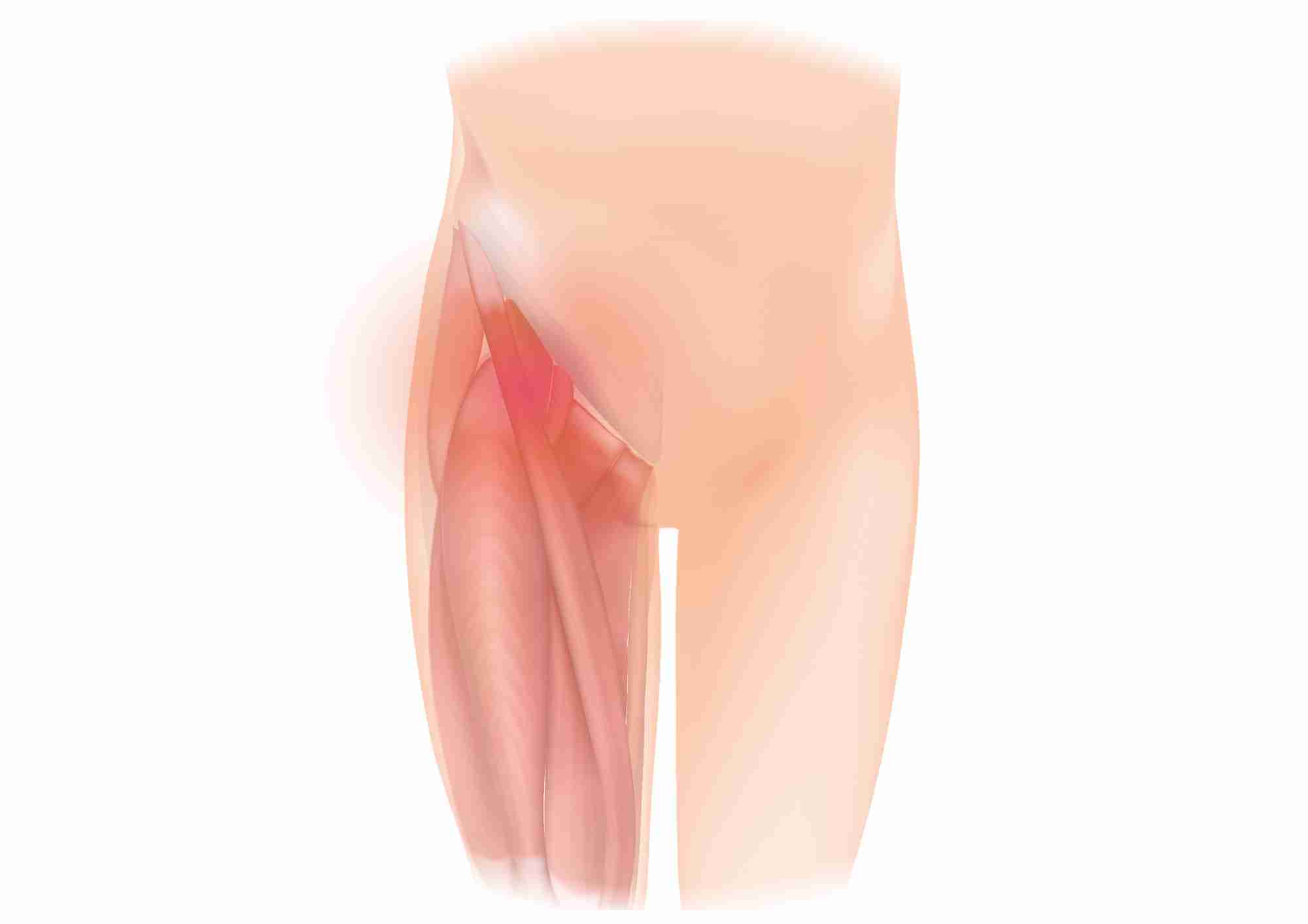
Distal Quadricep
Pain Relief
Inflammation Reversal
Collagen Synthesis
Shockwave Therapy for Distal Quadricep
Welcome to Houston Shockwave, where we offer advanced and effective shockwave therapy for distal quadriceps injuries. If you are experiencing pain or discomfort in the front of your thigh, you may have a distal quadriceps injury that could benefit from our cutting-edge treatment.
The quadriceps muscles are located at the front of the thigh and are responsible for extending the knee. The distal quadriceps, specifically, refer to the area where the muscle connects to the knee cap. When these muscles are stretched beyond their limits or subjected to repetitive stress, they can become strained or even torn.
The symptoms of a distal quadriceps injury include pain in the front of the thigh, swelling, and bruising. The severity of the injury can range from mild strains to complete tears, and the symptoms can range from mild discomfort to severe pain and difficulty walking or standing.
At Houston Shockwave, we offer non-invasive, safe, and effective treatment for distal quadriceps injuries using shockwave therapy. Shockwave therapy is a cutting-edge technology that uses high-frequency sound waves to stimulate healing and reduce pain in the affected area.
During a shockwave therapy session, a handheld device is placed on the skin over the affected area. The device then emits high-frequency sound waves that penetrate deep into the tissue, stimulating the body’s natural healing process.
Shockwave therapy has been shown to be effective in treating distal quadriceps injuries by promoting the regeneration of damaged tissue, increasing blood flow to the affected area, and reducing inflammation and pain. It is a non-invasive, outpatient procedure that requires no anesthesia or recovery time.
The benefits of shockwave therapy for distal quadriceps injuries include:
Non-invasive: Shockwave therapy is a non-invasive procedure that does not require surgery or injections.
Safe: Shockwave therapy is a safe treatment that has been used successfully for decades to treat a wide range of conditions.
Effective: Shockwave therapy has been shown to be effective in treating distal quadriceps injuries, with many patients experiencing significant pain relief and improved mobility after just a few sessions.
Quick and convenient: Shockwave therapy sessions typically last between 15 and 30 minutes, and patients can return to their normal activities immediately afterward.
Cost-effective: Shockwave therapy is a cost-effective alternative to more invasive treatments such as surgery or injections.
At Houston Shockwave, we have a team of experienced professionals who can help you determine if shockwave therapy is the best option for your specific needs. We will work with you to develop a personalized treatment plan that is tailored to your individual needs and goals.
Our team will also provide you with information and guidance on how to manage your symptoms and prevent further injury in the future. We believe that education is an important part of the healing process, and we are committed to helping our patients achieve optimal health and wellness.
If you are suffering from a distal quadriceps injury, shockwave therapy may be the right treatment for you. Contact Houston Shockwave today to schedule a consultation and learn more about how shockwave therapy can help you get back to a pain-free and active lifestyle. Don’t let a distal quadriceps injury hold you back any longer.

