281-357-5454

Houston Shockwave Therapy

Healing for chronic and acute injuries

Peroneal Nerve

Pain Relief
Inflammation Reversal
Collagen Synthesis

Pain In Your Foot?

Newest Techniques in Science. Non-Surgical.

Houston, TX Area

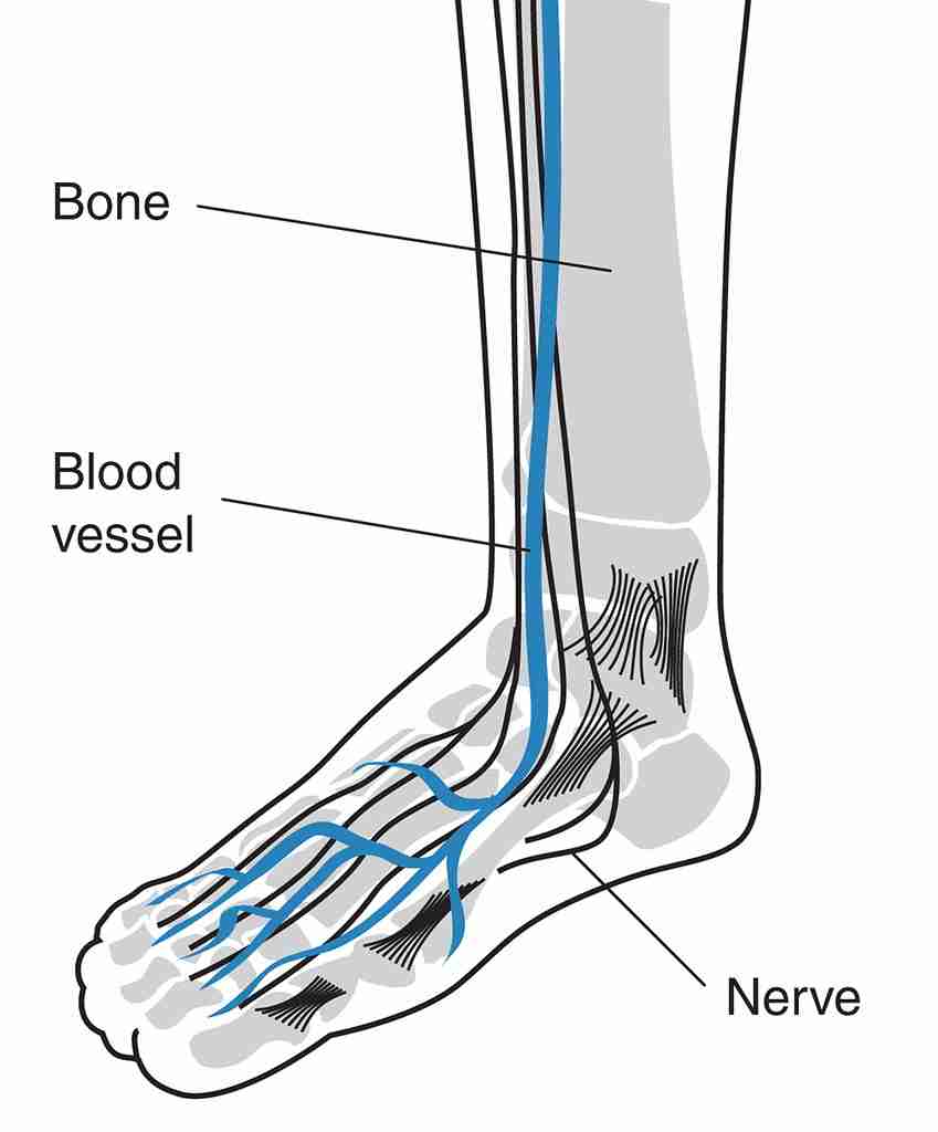
Shockwave Therapy for Peroneal Nerve

The common peroneal nerve usually referred to as the typical fibular nerve, is a significant nerve that innervates the lower extremity. It is one of both significant branches off the sciatic nerve as well as obtains fibers from the posterior divisions of L4 with S2 nerve roots. The common peroneal nerve divides from the sciatic nerve in the distal back thigh proximal to the popliteal fossa. After branching off of the sciatic nerve, it proceeds down the thigh, running posteroinferior to the arms femoris muscular tissue, as well as crosses laterally to the head of the lateral gastrocnemius muscle through the posterior intermuscular septum.

The nerve after that curves around the fibular neck prior to splitting right into two branches, the shallow peroneal nerve (SPN) and the deep peroneal nerve (DPN). The usual peroneal nerve does not have any kind of electric motor innervation prior to separating; however, it supplies sensory innervation to the lateral leg by means of the lateral sural nerve.

The superficial peroneal nerve innervates the side area of the leg, and also the deep peroneal nerve innervates the anterior compartment of the leg and the dorsum of the foot. These two nerves are vital in the eversion of the foot and dorsiflexion of the foot, specifically. The superficial peroneal nerve supplies both motor as well as sensory innervation. The innervation of the shallow peroneal nerve appears listed below:

Electric motor

Side compartment

Peroneus longus

Peroneus brevis

Sensory

Anterolateral leg


The deep peroneal nerve likewise gives both motor and sensory innervation in the leg. Deep peroneal nerve innervation is as complies with: 

Motor

Anterior area

Tibialis former

Extensor hallucis longus

Extensor digitorum longus

Peroneus tertius

Sensory

First dorsal 


Typical peroneal nerve: lateral facet of the leg just distal to the knee

Shallow peroneal nerve: the anterolateral distal third of the leg and also the majority of the dorsum of the foot (except the first webspace).

Deep peroneal nerve: first dorsal webspace.

Electric motor.

Usual peroneal nerve: none.

Superficial peroneal nerve: eversion of the foot.

Deep peroneal nerve: dorsiflexion of the foot as well as fantastic toe extension.

This paper will concentrate on peroneal nerve injuries, including etiologies, epidemiology, background, and physical exam searching for, in addition to diagnosis and treatment.


Etiology

There have been numerous stressful and atraumatic sources of peroneal nerve injury. Common causes of damage to the peroneal nerve consist of the following:.

Trauma or injury to the knee.

Knee dislocation.

Typical peroneal nerve injury can take place in up to 40% of patient.

A straight effect, penetrating injury, or laceration.

Injury tendency as the nerve training courses around the fibular head/neck.

Fracture of the fibula, especially at the proximal fibula.

Anatomic risk elements.

Common peroneal nerve entrapment can happen second to a fibrous band at the beginning of the peroneus longus.

Outside compression resources.

Limited splint/cast.

Compression wrapping/bandage.

Regular leg crossing.

Long term bed remainder.

Can be seen in comatose clients or those intubated for prolonged periods.

Positioning throughout anesthetic and surgical treatment.

Important to always pad bony prestiges, consisting of the location of the fibular head and neck at the lateral aspect of the knee.

Intraneural ganglion.

Peripheral nerve tumor.

Iatrogenic injury following surgery about the hip, knee, and ankle joint.

The typical peroneal nerve is most at risk to injury as it circulates the fibular neck. As a result it is most prone to injury at the side aspect of the knee.


The general incidence of peroneal nerve injury second to closed cracks is challenging to elucidate, provided the absence of prospectively acquired information. Fractures of the shin (including tibial plateau) and/or fibula are reportedly related to about a 1 to 2% rate of peroneal nerve injury. There are records of also rarer occurrences complying with overall knee arthroplasty (TKA) or arthroscopic lateral meniscal fixing.

Considerable injury around the knee can likewise result in peroneal nerve injury as a result of the nerve’s distance to the knee joint itself as well as its superficial place; this can consist of a straight effect to the fibular neck, lacerations, as well as knee misplacements. Typical peroneal nerve injuries are commonly encountered in athletes, particularly football or football players, either in association with knee dislocation and also ligamentous injury or alone.


Numerous systemic ailments can create compressive peroneal neuropathy and injury to the usual peroneal nerve, including:.

Diabetes mellitus.

Inflammatory problems.

Motor neuron illness.

Anorexia (exceedingly slim states) because of loss of subcutaneous fat at this degree causing compression of the nerve.


Epidemiology

Typical peroneal neuropathy is the commonest mononeuropathy experienced in the reduced limbs and also the 3rd most usual focal neuropathy encountered on the whole, after typical (repetitive strain injury) as well as ulnar neuropathies. Traumatic injuries to the usual peroneal nerve (CPN) most commonly affect young sports individuals (e.g., football, soccer) and grown-up patients complying with high power trauma (e.g., automobile crashes (MVCs)), with CPN injury supposedly occurring in 16 to 40% of individuals adhering to knee misplacements. Reduced power knee misplacements can happen in obese clients throughout basic tasks of day-to-day living.


History & Physical

A detailed history and physical exam to assess the standing and feature of the usual peroneal nerve is constantly needed. In individuals with a peroneal nerve injury, professional presentation varies based on the area and also extent of the injury as well as the visibility of anatomic variations. One of the most usual presentation of a common peroneal nerve injury is a weak point of ankle dorsiflexion and the traditional resultant foot decrease or capturing the toes while ambulating. Foot decrease can develop acutely or over days to weeks, depending upon the etiology. It can also be total or partial in extent. There might also be going along with numbness or paresthesia existing along the lateral leg, dorsal foot, and/or the first toe webspace. Discomfort may likewise be present in terrible situations but is not always present.


The clinician guides the clinical physical exam by the patient’s reported signs and symptoms as well as needs an understanding of the relevant anatomy and also possible underlying etiology. As an example, in cases of believed persistent peroneal nerve palsy with resultant foot decrease, the clinician ought to consider a main documented part of the assessment focusing on stride analysis as it may supply significant clues to the etiology of the signs and symptoms. An individual with damaged or paralyzed dorsiflexors might ambulate with a high steppage gait to stop dragging their toes on the ground due to nerve injury. Furthermore, tingling or dysesthesia in the upper lateral leg shows a lesion proximal to the fibular head, which may involve the sciatic nerve or lumbosacral nerve roots. In contrast, if there is a decreased or abnormal feeling in the lower side leg as well as dorsum of the foot, the surface peroneal nerve might be entailed. If there is additionally modified feeling in the dorsal facet of the very first internet area of the foot, the deep peroneal nerve might likewise be involved.


In the setting of traumatic injuries to the knee, including knee dislocations, the initial exam testing is done to eliminate an acute neurovascular injury that could potentially endanger the reduced extremity. Knee dislocations can notoriously jeopardize the neurovascular structures passing from the upper leg and also knee to the reduced arm or leg and also, otherwise dealt with urgently, can lead to severe compartment syndrome (AIR CONDITIONER), which can cause long-lasting compromise to the lower arm or leg calling for operative amputation.


To evaluate for the motor involvement of the shallow peroneal nerve as well as deep peroneal nerve, one have to examine foot eversion (SPN) and also foot/toe dorsiflexion (DPN). A finding of weak point of both foot eversion as well as foot/toe dorsiflexion recommends a lesion involving the usual peroneal nerve. Proximal lesions, for example, second to terrible knee misplacements, will certainly typically provide with varying levels of pins and needles in both superficial peroneal nerve and deep peroneal nerve circulations. A cautious and comprehensive assessment detecting the existence or absence of dorsiflexion of the ankle joint and/or terrific toe past neutral is necessary for electric motor evaluation testing. The refined difference is important in differentiating the ability to proactively dorsiflex the great toe from a maximally plantarflexed placement to a near-neutral setting. Such searchings for do not rule out deep peroneal nerve injury.


Tinel indicator is additionally a trusted scientific indicator to center the location of nerve irritation or entrapment. The examiner carries out the Tinel test by touching along the program of the nerve– especially around the fibular neck. If tingling or paresthesia is elicited distally to the point of compression by tapping, it signifies a favorable Tinel sign.


Examination

When the background as well as checkup is indicative of a prospective injury to the typical peroneal nerve, ordinary radiography should become part of the initial workup. Because of the proximity of the common peroneal nerve to the fibular neck along with its surface place, it is specifically vulnerable to guide trauma injury along with entrapment by soft as well as bony cells. Along with simple radiography, CT scans can be utilized to review more bony abnormalities, and also MRI/ultrasound can be used to assess soft-tissue resources or masses. The previous ought to be taken into consideration in the setting of tibial plateau cracks, while the last (i.e., MRI) is prudent in cases of traumatic knee dislocations.


Electrodiagnostic researches, including nerve conduction rate (NCV) examinations as well as electromyography tests (EMG), can be made use of to diagnose peroneal nerve palsy. These tests aid in the analysis of the motor and sensory axons of the peroneal nerve and also its branches. They are also handy in the localization of nerve injury. These examinations are useful in people offering with new-onset of peroneal nerve symptoms such as foot drop without a distressing mechanism along with assessing clients in the postoperative setting of a recognized stressful peroneal nerve injury to plan long-term administration as well as to give individual.


How shockwave therapy treatment can help:

New blood vessels are developed, improving blood supply and also oxygenation of the treated area. Faster healing is one result.
Reversal of persistent inflammation happens as pole cell activity rises. Pole cells play an important function in injury recovery and protection versus virus.
Accelerates the generation of collagen, which assists tendon fibers come to be denser and stiffer, producing a stronger structure.
Calcium build-up breaks down– granular portions of the buildup are eliminated by the body’s lymphatic system in areas where micro-tears and various other trauma to ligaments exist.
Material P levels are decreased and also discomfort is reduced when a natural chemical passes on pain messages to the central nerve system and inhibits the development of inflammatory edema.

Give us a call today and get on your path to recovery with Houston Shockwave Therapy located in The Woodlands, TX.


P: 281/357-5454

www.houstonshockwave.com