281-357-5454

Houston Shockwave Therapy

Healing for chronic and acute injuries

Hamstring Injuries

Pain Relief
Inflammation Reversal
Collagen Synthesis

Back of Leg Hurting?

Newest Techniques in Science. Non-Surgical.

Houston, TX Area

Shockwave Therapy for Hamstring Injuries

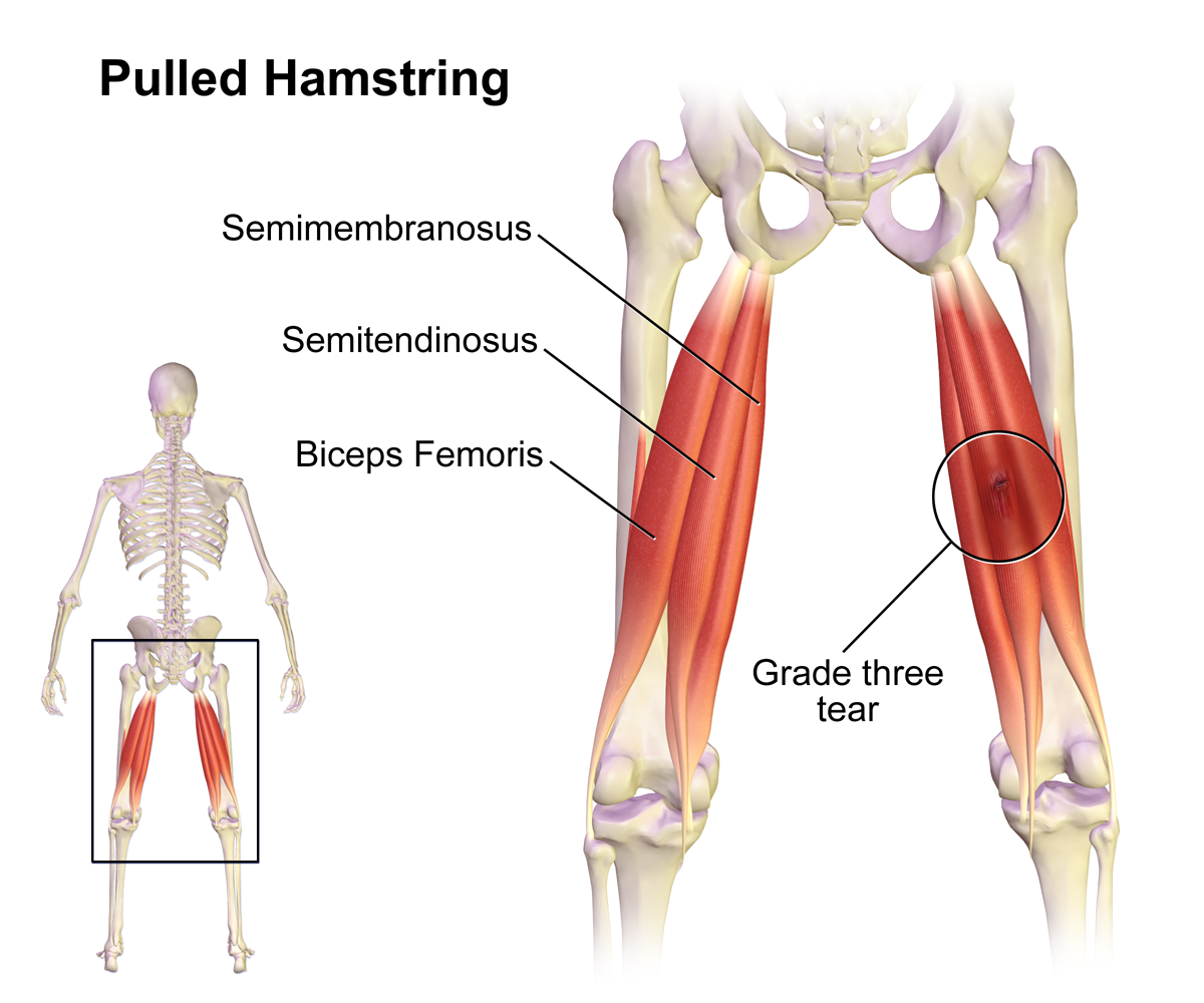
Hamstring muscles make up the three major muscular tissues of the posterior of the thigh. These consist of the semi-membranous in the inside of the leg, biceps femoris as the outermost, and semitendinosus in between. These teams of muscles are medically substantial as they are extremely prone to injury, particularly in athletes. All the hamstring muscular tissues pass through two joints (hip and knee) from beginning to insertion except the short head of biceps femoris, which only goes across knee joint from its origin to insertion. The primary feature of the hamstring muscles is flexion at the knee joint as well as extension at the hip joint. Biceps femoris additionally aids in the exterior turning of the hip while semimembranosus as well as semitendinosus help in the interior turning of the hip joint.

Injuries mostly happen while players are running. These teams of muscles are specifically at risk of injury due to their structural plan. Additionally, their system of activity over two joints (knee and hip) take place together with opposing effects on the hamstring length, which makes them susceptible to injury. Their essential role in slowdown while walking, running, and making acute changes in direction, makes them much more susceptible to being injured. Hamstrings bear the significant strain during the stage of motion when they shift from slowing down the expansion of the knee to prolonging the hip joint. During this quick shift of functional biomechanics, they are most at risk to injury.


Grading of Injury

Quality 1: Light pain or swelling, non-palpable tissue disruption, none or minimal loss of capacity.

Grade 2: Moderate pain and swelling, bringing about loss of capacity.

Grade 3: Total interruption or tear of the musculo-tendinous unit with severe discomfort and swelling and lack of function.


There have actually been some researches done to highlight the etiologies and threat aspects related to a hamstring injury. All the research studies tend to suggest hamstring injury has a multifactorial origin. Interaction amongst different danger elements is of paramount value when taking into consideration the origin of this sort of muscle injury. A previous hamstring injury, older age, and peak quadriceps torsion are highly related to the risk of a hamstring injury. Ethnic culture, specifically African and Indigenous origin and a higher degree of competitiveness, have also been found to be connected with a higher occurrence of hamstring injury. Other threat variables like hamstring versatility, the weight of the athlete, hip flexor, quadriceps versatility, and hamstring peak torsion require further study to be proven as potential danger factors.


The British Medical Journal published a short article with a research study carried out in 100 soccer players, which revealed practical asymmetry of the muscular tissues inclined them to a greater risk of sustaining a hamstring injury. Additionally, the occurrence of hamstring injury is, the majority of the time, sports related. Sports that need quick velocity and slowdown like American football, soccer, and rugby had much more occurrence of a hamstring injury. More extreme injuries have actually been located through kicking, while a greater incidence of injuries happens during running. Similarly, high-intensity training sessions, a higher degree of competition and positions in play that require more operating are associated with more hamstring injuries.


Research studies showed hamstring injury peaks at 16 to 25 years old. It is primarily related to sporting activities where hamstrings bear the most responsibility, with a fast change of its practical function while in use like running, football, soccer, track and field sports. One NFL research done over a decade showed greater than 50% injury throughout the preseason with mainly defense and pass receivers, that demand more speed.


When an athlete develops an acute posterior thigh discomfort through a match, which makes it tough to go back to play, a hamstring injury should be presumed. Intense hamstring injuries have pain with weight bearing and an irregular stiff-legged gait as a result of staying clear of hip and knee flexion. If there is a complete break from the ischial tuberosity, pain can be localized to the proximal upper leg with point inflammation at the ischial tubercle. Individuals commonly have difficulty resting as a result of discomfort at the site. This clinical presentation is called gluteal sciatic nerve pain. It is related to compression of the sciatic nerve. Knee flexion could be affected as well.


Checkup, though a vital part of the preliminary evaluation of hamstring injury, must not be relied upon completely to identify the precise nature of the injury due to the deep area of the hamstring muscle. Local swelling in the posterior upper leg with tenderness to palpation and hardness in the posterior upper leg along the hamstring muscular tissue bellies are the typical assessment areas. Full tear might have an apparent flaw within the muscular tissue make-up. At the time of physical exam, strength, and range of activity of the affected hamstring needs to be evaluated and compared to those on the unaltered side. Hip flexion and knee expansion ought to be taken a look at, which may be restricted by discomfort in patients with a hamstring injury.


Imaging is a crucial facet of the evaluation of possible hamstring injury to establish the nature, extent, and seriousness of the injury. Ultrasound and MRI are the diagnostic methods of option. Area of injury, as shown by edema and hemorrhage, is depicted by echotexture in ultrasound as well as high signal intensity. Ultrasound allows vibrant analysis of the affected hamstring muscle; however, its sensitivity is enhanced if the ultrasound is done early after injury. MRI is still the imaging of choice to evaluate much deeper muscular tissue injuries, assess a recurrent injury, and to separate new injury from the residual scarring. MRI imaging also gives a quote of the recovery duration. Though it can predict projected time far from the sport, it cannot identify the threats for re-injury, which needs further study.


How shockwave therapy treatment can help:

New blood vessels develop, improving blood supply and oxygenation of the treated area. Reversal of persistent inflammation happens as pole cell activity rises. Pole cells play an important function in injury recovery and protection versus virus. Accelerates the generation of collagen, which assists tendon fibers to be denser and rigid, producing a stronger structure. Calcium build-up breaks down– granular portions of the buildup are eliminated by the body’s lymphatic system in areas where micro-tears and various other trauma to ligaments exist. Material P levels are decreased and discomfort is reduced when a natural chemical passes on pain messages to the central nerve system and inhibits the development of inflammatory edema.

Give us a call today and get on your path to recovery with Houston Shockwave Therapy located in The Woodlands, TX.


P: 281/357-5454

www.houstonshockwave.com